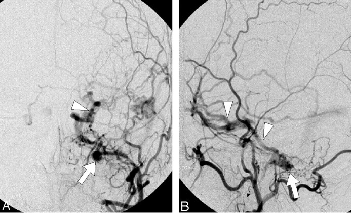
Fig 7.
Frontal (A) and lateral (B) views of preoperative external carotid arteriography. The dural arteriovenous fistula is fed by meningeal branches of the ascending pharyngeal artery and occipital artery. A dilated venous sac is seen at the fistulous point (arrow). Note the retrograde venous drainage via the inferior petrosal sinus and cavernous sinus (arrowheads).

