
Fig 6.
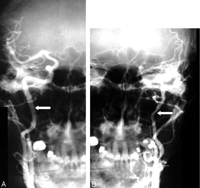
Decreased diameter of the ipsilateral ICA compared with the normal opposite carotid artery.
A, Anterioposterior right carotid angiogram shows the diameter of the normal ICA (arrow).
B, Anterioposterior left carotid angiogram done with the same magnification (same position of radiograph tube and image intensifier; same field of view) as the right in A showing decreased diameter of the left ICA (arrow).