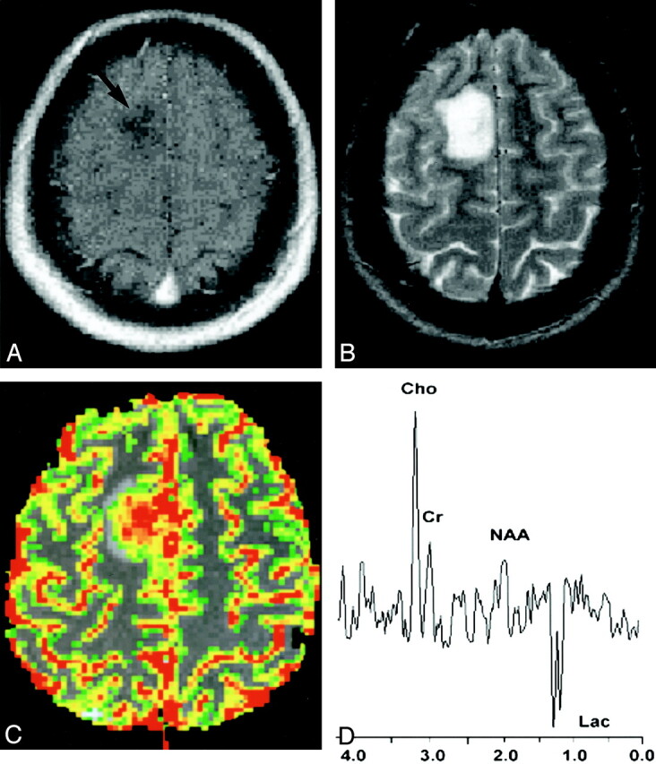
Fig 1.

20-year-old woman with biopsy-proved high-grade glioma.
A, Contrast-enhanced axial T1-weighted image (600/14/1 [TR/TE/NEX]) demonstrates an ill-defined nonenhancing mass (arrow) in the right frontal region. The lack of enhancement on the conventional MR image suggests a low-grade glioma.
B, Axial T2-weighted image (3400/119/1) shows increased signal intensity in the mass, with minimal peritumoral edema. This mass was graded as a low-grade glioma with conventional MR imaging because of lack of enhancement, minimal edema, no necrosis, and no mass effect.
C, Gradient-echo (1000/54) axial perfusion MR image with rCBV color overlay map shows increased perfusion with a high rCBV of 7.72, in keeping with a high-grade glioma.
D, Spectrum from proton MR spectroscopy with the PRESS sequence (1500/144) demonstrates markedly elevated Cho and decreased NAA with a Cho/NAA ratio of 2.60, as well as increased lactate (Lac), in keeping with a high-grade glioma.
