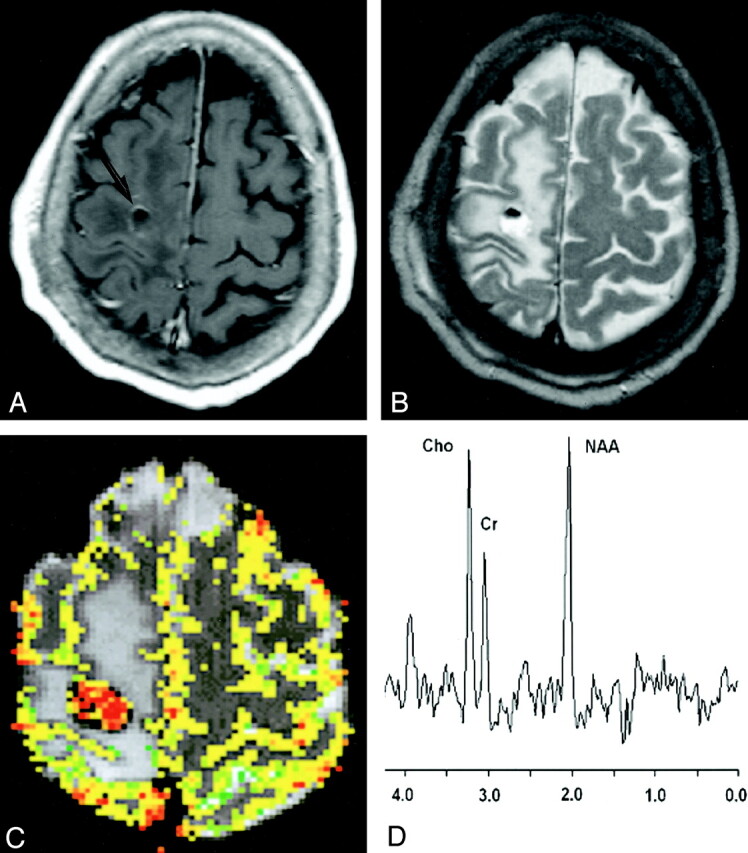
Fig 2.

43-year-old man with biopsy-proved low-grade glioma.
A, Contrast-enhanced axial T1-weighted image (600/14/1) demonstrates a peripherally enhancing mass (arrow) in the right frontal region. The presence of contrast material enhancement on the conventional MR image would suggest a high-grade glioma.
B, Axial T2-weighted image (3400/119/1) shows marked peritumoral edema with possible necrosis and blood products. This mass was graded as a high-grade glioma with conventional MR imaging because of the contrast material enhancement, heterogeneity, blood products, possible necrosis, and degree of edema.
C, Gradient-echo (1000/54) axial perfusion MR image with rCBV color overlay map shows a low rCBV of 1.70, in keeping with a low-grade glioma.
D, Spectrum from proton MR spectroscopy with the PRESS sequence (1500/144) demonstrates elevated Cho and slightly decreased NAA with a Cho/NAA ratio of 0.90, which is more in keeping with a low-grade glioma.
