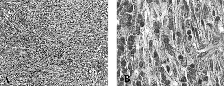
Fig 3.

Microscopic images.
A, Proliferation of spindle cells oriented in intersecting fascicles or haphazardly distributed, accompanied by numerous plasma cells and small lymphocytes (original magnification, ×200).
B, Myofibroblasts show minimal nuclear pleomorphism (original magnification, ×1000)
