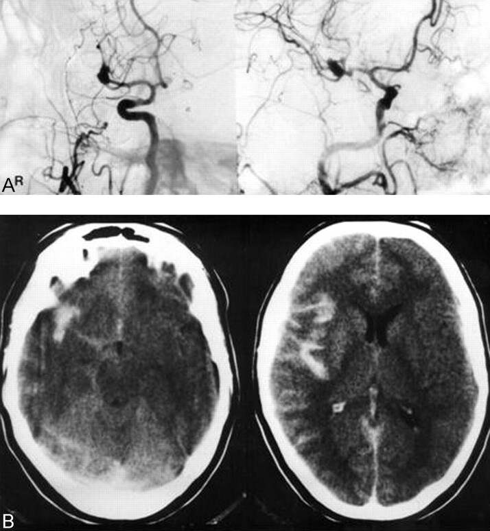
Fig 1.
Case 1. A, Right ICA angiogram (anteroposterior view [left] and lateral-oblique view [right], arterial phase) shows an MCA aneurysm. During angiography, blood pressure dropped and an episode of bradycardia and mild local vasospasm of proximal MCA branches developed without visible extravasation.
B, CT scans obtained immediately after angiography shows an SAH located in the right sylvian fissure.

