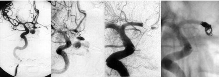
Fig 3.
Right ICA angiogram, anteroposterior view. Active bleeding from the reruptured anterior communicating artery aneurysm during the angiography is visible (left two pictures). The dome of the aneurysm is completely lacerated. Extravasation of contrast material into the chiasmal cistern is visible. The diameter of the neck is 1.9 mm, and the diameter of the fundus 4.5 mm. After detachment of the first coil, bleeding continued, and protrusion of the coil into the subarachnoid space was imminent. Closure of the aneurysm with parent vessel occlusion of the A1-A2 segment of the right anterior cerebral artery (right two pictures).

