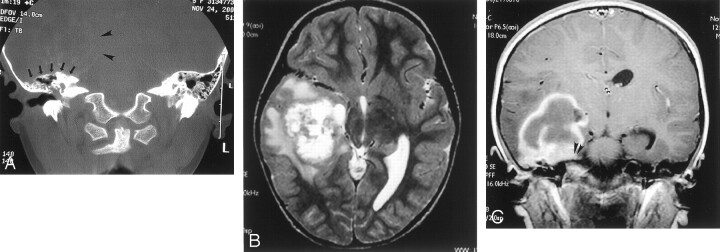
Fig 1.
Images from the case of a 5-year-old left-handed girl who presented with a 2-month history of intermittent headache that was associated with nausea and vomiting during the week before diagnosis.
A, Coronal CT bone window scan shows a large, ill-defined, intra-axial mass located in the right temporal lobe (arrowheads) with remodeling of the tegmen tympani (arrows) but no direct invasion of the temporal bone.
B, Axial T2-weighted MR image shows a heterogeneous mass in the right temporal lobe.
C, Coronal contrast-enhanced T1-weighted MR image shows a heterogeneously enhancing mass with mild dural enhancement (arrowheads) but no extension into the underlying petrous temporal bone.

