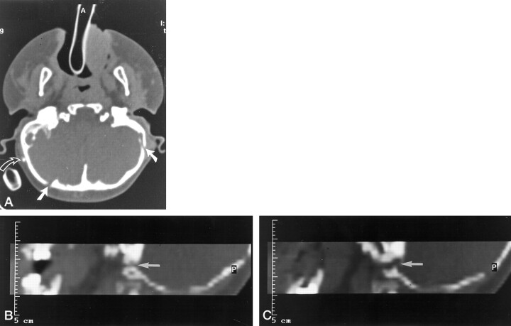
Fig 3.
A, Axial CT scan (study group, patient 5) shows bilateral jugular foramen stenosis and enlarged transosseous emissary foramina in a case of Apert syndrome. The patient has a ventriculoperitoneal shunt (curved arrow).
B and C, Parasagittal reformatted CT scans of the same patient show narrowed and tortuous jugular foramina (arrows) (compare with Fig 4).

