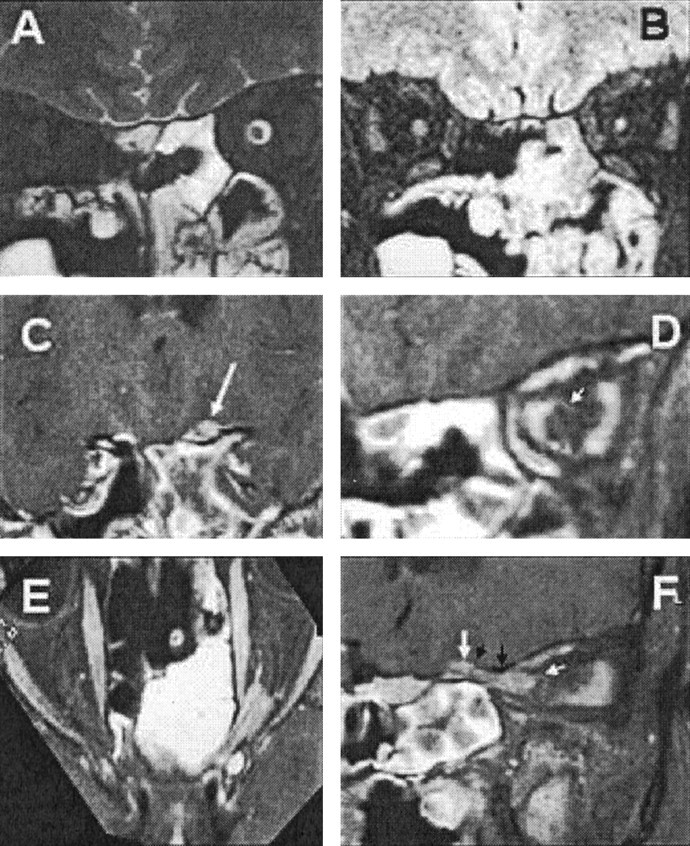
Fig 2.

Case 3.
A, T2-weighted coronal MR image, showing dilatation of the left optic nerve sheath.
B, Heavily T2-weighted image with fat and water suppression (SPIR/FLAIR), showing increased T2 signal intensity and atrophy of the optic nerve.
C, Coronal images through the sulcus chiasmaticus, demonstrating a small tumor nodule arising from the cranial end of the left optic canal (arrow).
D, Coronal image from a contrast-enhanced T1-weighted volume-rendered MR image, acquired with fat suppression through the optic nerve in the midorbit, showing en-plaque growth of tumor within the left optic nerve sheath (arrow).
E, Axial oblique reconstruction from a contrast-enhanced T1-weighted volume-rendered MR image, acquired with fat suppression along the optic canals, showing the extent of the tumor. Note the optic nerve emerging from the tumor anteriorly and the en-plaque growth of tumor along the anterior optic nerve sheath.
F, Sagittal oblique reconstruction from a contrast-enhanced T1-weighted volume-rendered MR image, acquired with fat suppression along the course of the left optic canal, showing the extent of the tumor. Note the tram track sign due to the central nonenhancing optic nerve, the mural nodule (large white arrow) arising behind the roof of the optic canal (black arrows), and the en-plaque growth of the tumor along the anterior part of the orbital optic nerve sheath (small white arrow).
