Fig 1.
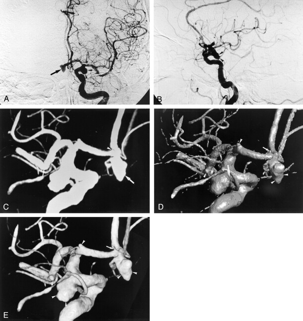
Multiple aneurysms (of one anterior communicating artery, two anterior cerebral arteries, one middle cerebral artery, and one posterior communicating artery) in a 73-year-old woman.
A, Anteroposterior and B, lateral 2D DSA images of the left internal carotid artery depict a 7-mm anterior communicating aneurysm (arrow in A) and a 9-mm posterior communicating aneurysm (arrowheads). The other aneurysms are not clearly shown on the 2D DSA images. On rotational DA images (not shown), one 1.5-mm aneurysm at the A1 segment was not detected by the two observers.
C–E, Three-dimensional DA images from behind reveal the five aneurysms (of one anterior communicating artery, two anterior cerebral arteries, one middle cerebral artery, and one posterior communicating artery). The MIP 3D image (C) does not clearly demonstrate a 1.5-mm aneurysm (arrowhead) at the A1 segment and a 2.5-mm aneurysm (small arrow) at the M1-M2 segment, which was not detected by the observers. Delineation of the shape of the anterior communicating aneurysm (large arrow) on the MIP 3D image is inferior to that on the SSD (D) and volume-rendering (E) 3D images because of lack of depth. On the SSD 3D image, the neck size of the posterior communicating aneurysm (large arrow in D) is overestimated compared with that on the volume-rendering 3D image (E). It is not clear whether the very small aneurysm (small arrow in D) at the A1 segment is an aneurysm or not. The two observers ranked it as ambiguous visualization. The other three aneurysms (arrowheads) are clearly demonstrated. On the volume-rendering image, the four aneurysms (arrowheads in E) were classified as sufficient visualization. However, the very small aneurysm (arrow in E) at the A1 segment was evaluated as ambiguous visualization. All the aneurysms were proved at surgery.

