
Fig 5.
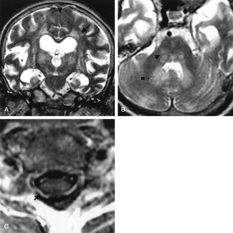
Patient 5. Paralytic rabies.
A, Coronal and B, axial fast spin-echo T2-weighted images of the brain, and C, gadolinium-enhanced axial T1-weighted image of the midcervical cord reveal ill-defined moderate hyperintensity changes of the deep gray matter (double arrows in A), white matter (single arrow in A), and brain stem and dentate nuclei (arrowhead and arrow with block in B). Vivid enhancement of the ventral and dorsal cervical nerve roots (arrows in C) is demonstrated. (Reprinted with permission from reference 3.)