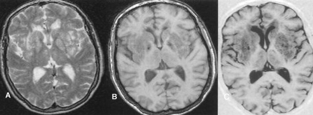
Fig 1.
Matching axial MR images show severe (grade 5) dilatation of the VRS (matrix = 256 × 256, FOV = 230 × 230 mm): A, T2-weighted variable echo (TR/TE1/TE2 = 5500/20/90); B, T1-weighted high-spatial-resolution T1-weighted 3D gradient echo (TR/TE = 24/18, section thickness = 0.89 mm, flip angle = 30°); and C, inversion recovery (TR/TE/TE = 6850/18/300). Calculated CNRs for VRS versus WM are 64.1 for inversion recovery, 24.8 for fast field-echo, 19.1 for the variable-echo second-echo imaging.

