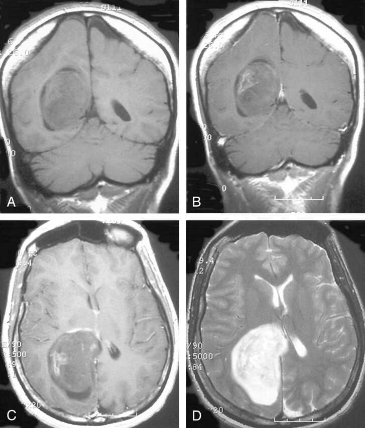
Fig 2.

Precontrast coronal (A), postcontrast coronal (B), postcontrast axial (C), and T2-weighted axial (D) MR images redemonstrating a minimally enhancing mass within the trigone. The mass is homogenous and well-defined and appears to cause local expansion of the ventricle.
