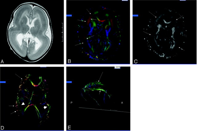
Fig 1.
Patient, a 30-day-old male with isolated lissencephaly.
A, Axial T2-weighted image shows diffuse thickening of the cortex with lack of sulcation.
B, Axial 2D color map through the body of the lateral ventricles shows bands of color (arrows) corresponding to the deep cortex.
C, Axial anisotropy map shows bands of higher anisotropy corresponding to the bands of color (arrows) seen on the color maps.
D, Axial 3D presentation of corpus callosum, fronto-occipital fasciculus (short arrows), and presumed cell layer IV (arrows).
E, Sagittal 3D depiction of the cingulum (long arrow) and fornix (short arrow). Note absence of the temporal projections of the cingulum and fornix.

