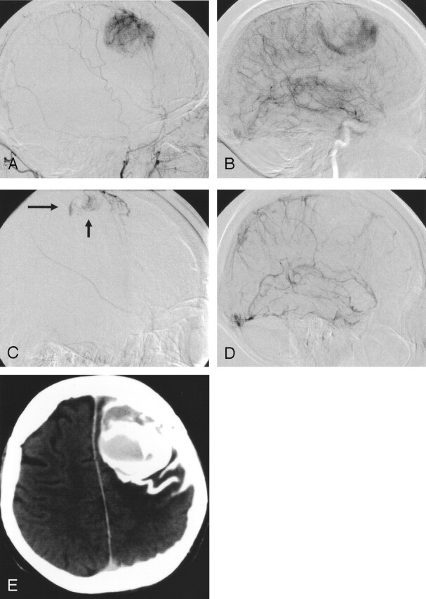
Fig 2.

Patient 11. Subarachnoid and intratumoral hemorrhage during embolization (Embospheres, 100–300 μm) of a right frontal-convexity meningioma.
A and B, Images show blood supply by the ipsilateral middle meningeal artery (A), which was subsequently devascularized with particles, and leptomeningeal branches of the middle cerebral artery (B).
C and D, At the end of the procedure, patient had sudden-onset headache. Angiograms show subarachnoid extravasation of contrast medium (arrows in C). Control run in the internal carotid artery (D) shows disappearance of the leptomeningeal supply, indicating complete tumor devascularization.
E, Postprocedural CT shows intratumoral and subarachnoid hemorrhage. At surgery, bleeding from intratumoral vessels were slight; the fresh intratumoral clot and tumor were easily removed. The patient recovered completely.
