Fig 3.

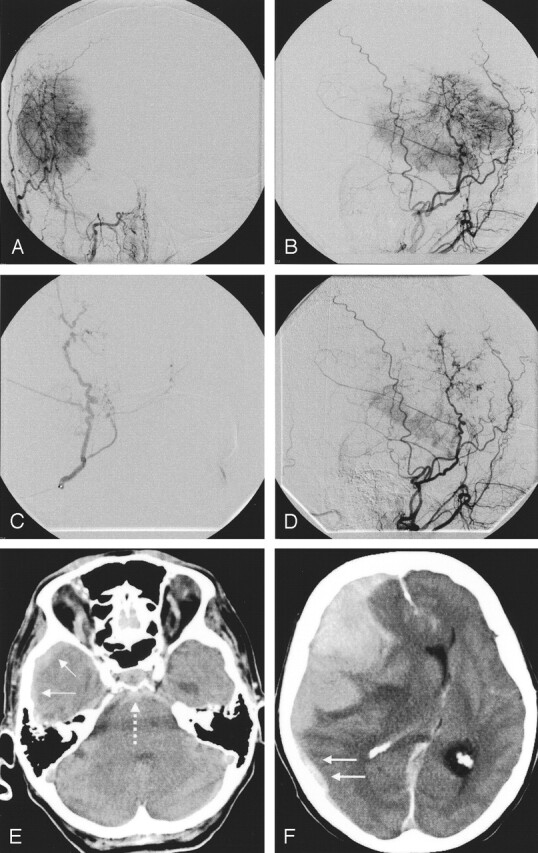
Patient 12. An 81-year-old woman with fatal subdural, subarachnoid, and intratumoral hemorrhages after embolization (Bead Block,100–300 μm).
A and B, Embolization of a large, right temporal meningioma with a predominant middle meningeal arterial supply.
C, Ipsilateral middle meningeal artery was superselectively probed and embolized with spherical particles.
D, Procedure was abandoned after the application of one vial because the patient had back pain. Control image reveals marked tumoral devascularization.
E and F, Afterward, the patient had no new neurologic symptoms, but 2 hours later, she was comatose with fixed, dilated pupils. CT shows extensive subdural (solid arrows), subarachnoid (dotted arrow) and intratumoral hemorrhage. Because of her age and clinical state, she did not undergo surgery and died the next day.
