
Fig 2.
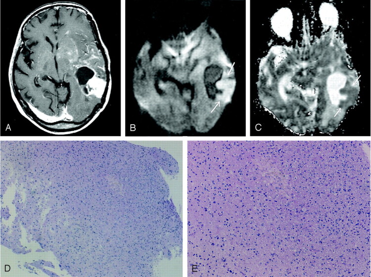
Images obtained in a 53-year-old woman with biopsy-proven tumor after receiving radiation and chemotherapy for anaplastic astrocytoma.
A, Gadolinium-enhanced T1WI image shows a ring-enhancing lesion with a solid enhancing component in the left temporal lobe. Multiple patchy enhancements with mass effect are also seen in the left basal ganglia and insula, suggestive of tumor infiltration.
B, DWI image obtained at the same level as that of A shows the solid enhancing component of predominant hyperintensity (arrows), which usually represents densely packed tumor cells.
C, ADC map, which shows the relatively low apparent diffusion coefficient value of the lesion (arrows).
D and E, Histopathological specimens (hematoxylin-eosin, ×20 [D], ×80 [E]) show tumor tissues with increased cellular density corresponding to anaplastic astrocytoma. No evidence of necrotic tissue was found.