Fig 3.
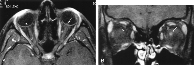
17-year-old girl with moderate imaging findings of optic neuropathy due to cat scratch fever (patient 4, Table 2).
A and B, Axial (A) and coronal (B) gadolinium-enhanced fat-suppressed T1-weighted images (TR1, TR2/TE, 735, 875/14) show bulging of the left optic disc (arrow, A) that is markedly less pronounced than that on the right (arrowhead, A) as well as that in Figure 1. The associated enhancement at the left optic nerve–globe junction (arrow, A and B) is also markedly less extensive. Note the normal appearance of the optic nerve–globe junction region on the right (arrowhead, A and B).

