
Figure 2.
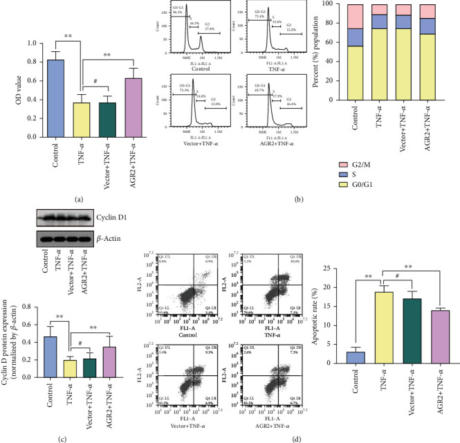
AGR2 overexpression promotes intestinal epithelial cell proliferation. (a) Viability of the Caco-2 cells as detected by CCK-8 assay. (b) The cell cycle distribution of the Caco-2 cells as determined by flow cytometry. (c) Cyclin D protein expression as determined by Western blot analysis. (d) The apoptosis level of the Caco-2 cells as determined by flow cytometry. The data are presented as the mean ± SD, #p > 0.05, ∗p < 0.05, ∗∗p < 0.01.