
Figure 3.
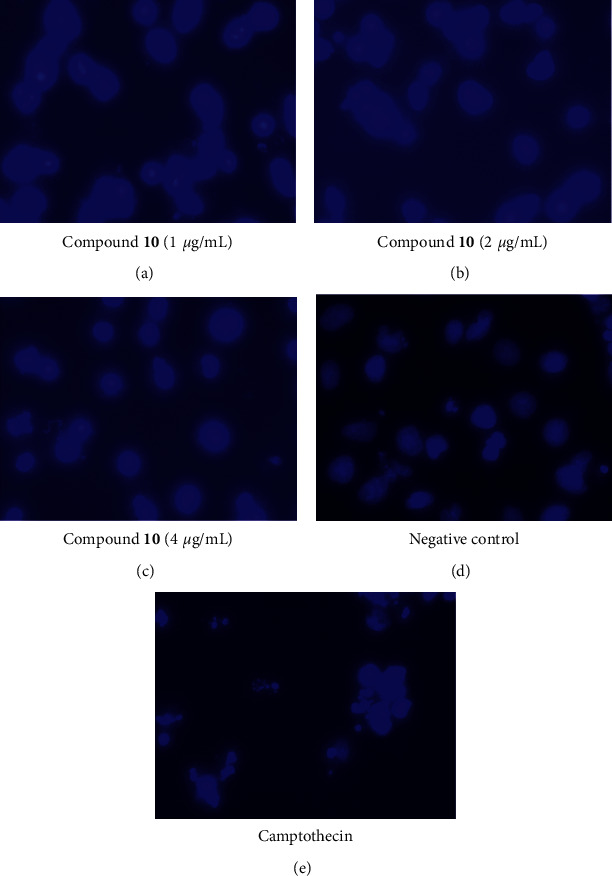
Nuclear condensation and fragmentation effects of compound 10 on NTERA cells at different concentrations as 1 µg/mL, 2 µg/mL, and 4 µg/mL using Hoechst 33342 staining. The cells were at 48 h of incubation and observed with Zeiss Vert A1 inverted microscope (100x).