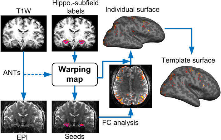
Figure 1.
The procedures of coregistration. Individual T1-weighted images were coregistered to the EPI by ANTs. The warping coefficients were then applied to the labeled images of the hippocampal subfields. The seed-based FC analysis was performed on the EPI space and the FC map was re-sampled onto individual cortical surface before being warped to the template surface. The re-sampling of FC values on EPI space captured only the signal from the white matter surface to the middle depth of cerebral cortex in order to suppress contamination from large vessels.

