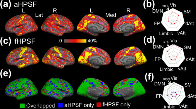
Figure 6.
Hippocampal coverage of iFC associated with aHPSF and fHPSF. (a) The cortical map of hippocampal coverage associated with aHPSF. The contours of functional networks are outlined. (b) The radar chart of averaged hippocampal coverage within each functional network associated with aHPSF. (c) The cortical map of hippocampal coverage associated with fHPSF. (d) The radar chart of averaged hippocampal coverage within each functional network associated with fHPSF. (e) The overlap between the binarized cortical maps in (a) and in (b). The overlapping area is highlighted in green. The area which connects only with aHPSF is highlighted in blue; fHPSF in red (f) The percentages of the overlapping, ‘aHPSF only’ and ‘fHPSF only’ area within each functional network. L left; R right; Lat lateral view; Med medial view; Vis visual network; SM sensorimotor network; dAtt dorsal attention network; vAtt ventral attention network; Limbic limbic network; FP frontoparietal network; DMN default-mode network.

