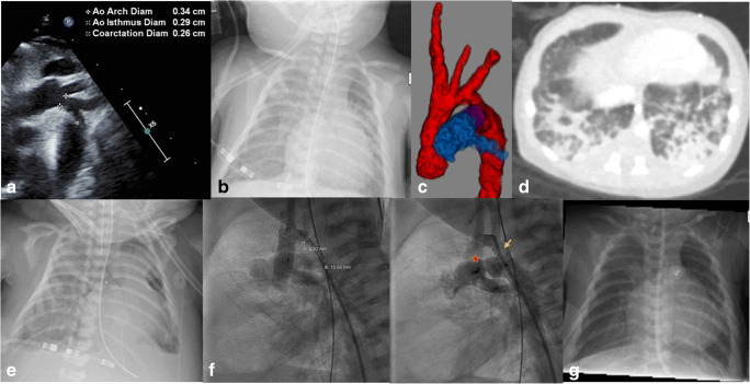
Fig. 1.
a ECHO showing coarctation. b Chest X-ray showing infiltrates. c CT angiography showing coarctation, hypoplastic aortic arch, and PDA. d “Ground glass” infiltrates in CT chest. e Chest X-ray after VA ECMO cannulation. f Chest fluoroscopy (arrow, ductal stent; star, PDA plug). g Chest X-ray post ECMO decannulation with resolution of infiltrates and vascular prostheses in situ. ECHO, echocardiogram; CT, computed tomography; PDA, patent ductus arteriosus; VA ECMO, venoarterial extracorporeal membrane oxygenation; ECMO, extracorporeal membrane oxygenation

