Figure 6.

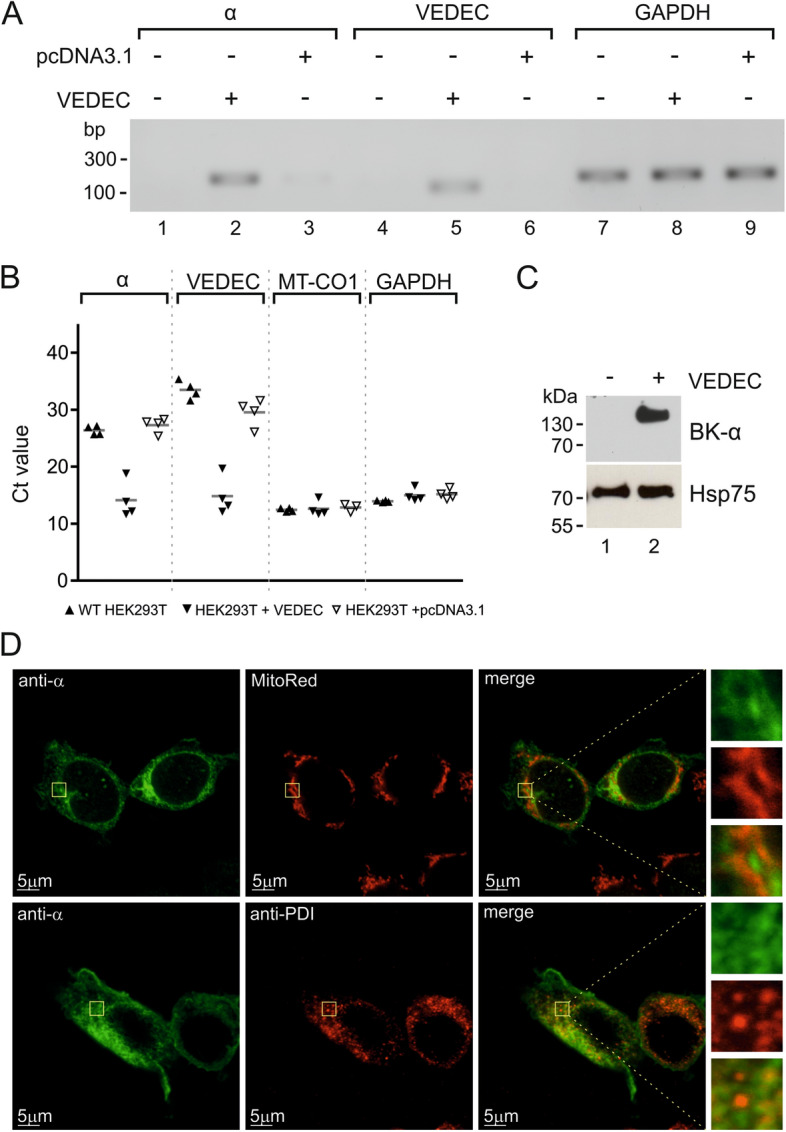
Analysis of the presence of the BKCa α subunit in wild-type and VEDEC transiently transfected HEK293T cells. (A) Analysis of selected gene expression using qualitative PCR. (B) Real-time PCR analysis of the expression levels of selected genes. (C) Western blot analysis of the BKCa pore-forming subunit in the mitochondrial fraction of wild-type and transfected cells. (D) Localization of VEDEC in HEK293T cells after transient transfection. Confocal images of cultured cells stained with MitoRed as a mitochondrial marker (upper panel, red channel) and an anti-PDI antibody as an endoplasmic reticulum marker (lower panel, red channel). The pore-forming α subunit was stained with an anti-BK α antibody (green channel). The superimposition of the two signals revealed the partial mitochondrial and ER localization of the BKCa α subunit (orange).
