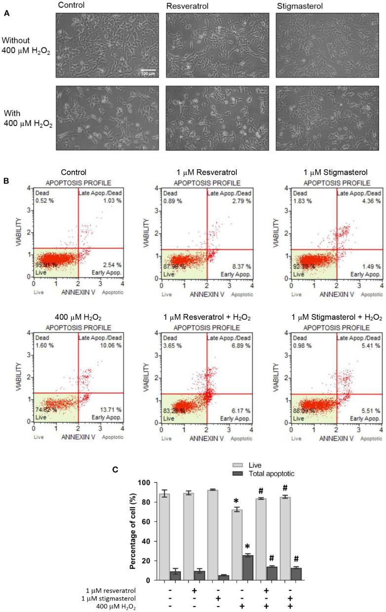
Figure 3.
Resveratrol and stigmasterol inhibited apoptosis caused by H2O2. (A) Morphology of SH-SY5Y cells was observed under the inverted-light microscope at 20 × magnification, scale bar 100 μm. (B) Representative plots of live and apoptotic cells as measured by flow cytometry on pretreatment of 1 μM resveratrol and stigmasterol exposed to 400 μM H2O2 for 24 h. (C) Bar chart of live and total apoptotic cells. Data are presented as mean ± S.E.M. of three independent experiments (*p < 0.05 vs. control; #p < 0.05 vs. H2O2-treated group).

