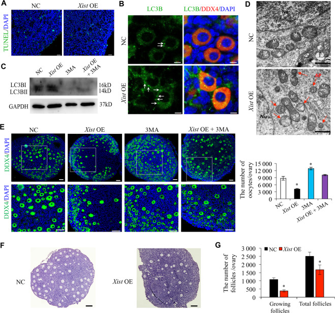
Fig. 2. Xist promotes oocyte autophagy during PF formation.
a TUNEL staining in newborn ovaries transfected with Xist-pcDNA3.1 (Xist OE) or the empty vector control (NC). The nucleus was stained by DAPI (blue). Scale bars: 20 μm. b Immunofluorescence staining of LC3B (green), and DDX4 (red) in newborn ovaries transfected with Xist-OE compared to NC control. Scale bars: 5 μm. Arrows indicating LC3B puncta. c WB analysis of active LC3B expression in newborn ovaries treated under indicated condition. d TEM analysis of autophagosomes in newborn ovaries transfected with Xist-OE compared to NC control. Red arrow indicating autophagosomes. Scale bars: 1 μm. e Representative images of DDX4 immunofluorescence staining (left) and quantification of follicles (right) in newborn ovaries treated under indicated condition. The nucleus was stained by DAPI (blue). Scale bars: 50 μm. f, g Representative images (f) and quantification of follicles (g) in the ovary transfected under indicated condition. Scale bar, 20 μm. Student’s t-test: *P < 0.05.

