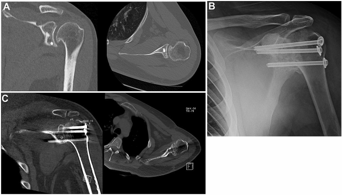
Fig. 2.
Case 2: a 25 year-old patient with recalcitrant post instability that after several failed stabilization procedures underwent GHA. a CT scan of the left shoulder showing coronal (left) and axial (right) projections of posterior screwed bone block procedure. Chronic instability led to early osteoarthritis with osteophytes in the infero-medial humeral head. b Radiograph of a left shoulder showing GHA using three cannulated screws with washers transfixing the glenohumeral joint. Fusion to the acromion was avoided. Post-operative CT scan of the left shoulder showing successful gleno-humeral fusion in coronal (left) and axial views (right)

