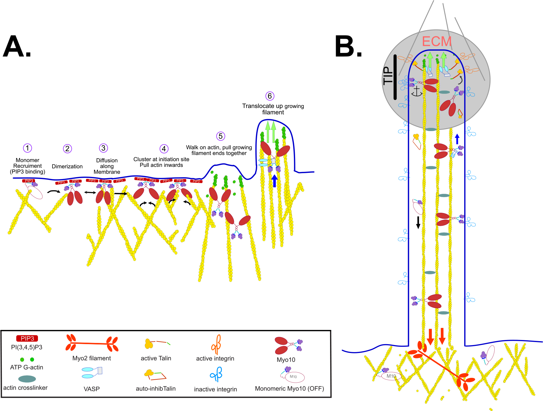
Figure 3. Model of MYO10 Function in Filopodia Formation and Function.

Schematic illustration of the activation and role of MYO10 in filopodia initiation, in transport of VASP towards the growing filopodia tip and in anchoring integrins at the filopodia tip. (A) MYO10 is a monomer prior to activation and binding to PI(3,4,5)P3 opens the molecule and promotes activation and dimerization (illustrated on the left side). Green arrows indicate extension of filopodium as monomers are added to the tip of the growing filopodium. Blue arrow represents myosin-driven transport of a cargo. (B) MYO10 within the filopodium can translocate along the actin core or return to the cytosol via retrograde flow generated by MYO2A. Red arrows indicate the downward pulling force exerted by MYO2A filaments in the cortex. ECM, extracellular matrix (highlighted with grey circle).
