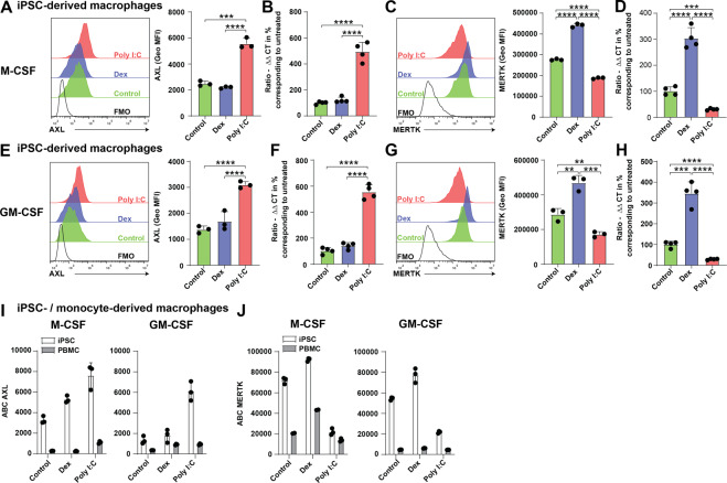
Fig. 1. Human iPSC-derived macrophages express AXL and MERTK.
A–H Macrophage progenitor cells were generated from iPSCs and cultured in vitro using either M-CSF or GM-CSF. Macrophages were stimulated with Poly I:C or dexamethasone or left untreated. After 24 h AXL and MERTK expression was determined by flow cytometry or by qRT-PCR. A–D Representative histograms showing expression of AXL and MERTK on M-CSF differentiated macrophages upon stimulation using indicated conditions. Bar graphs show geometrical mean fluorescence intensity of AXL and MERTK or mRNA expression levels of Axl and Mertk. Shown is mean with SD and individual samples. E–H Representative histograms showing expression of AXL and MERTK on GM-CSF differentiated macrophages upon stimulation with indicated conditions. Bar graphs show geometrical mean fluorescence intensity of AXL and MERTK or mRNA expression levels of Axl and Mertk. Shown is mean with SD and individual samples. I, J Absolute flow cytometric quantification of AXL and MERTK cell surface expression on iPSC- and monocyte-derived macrophages. Antibodies per cell (ABC) have been determined using a bead based assay. All experiments have been performed with three (flow cytometry) or four (qRT-PCR) technical replicates. Data were representative of at least three individual experiments.

