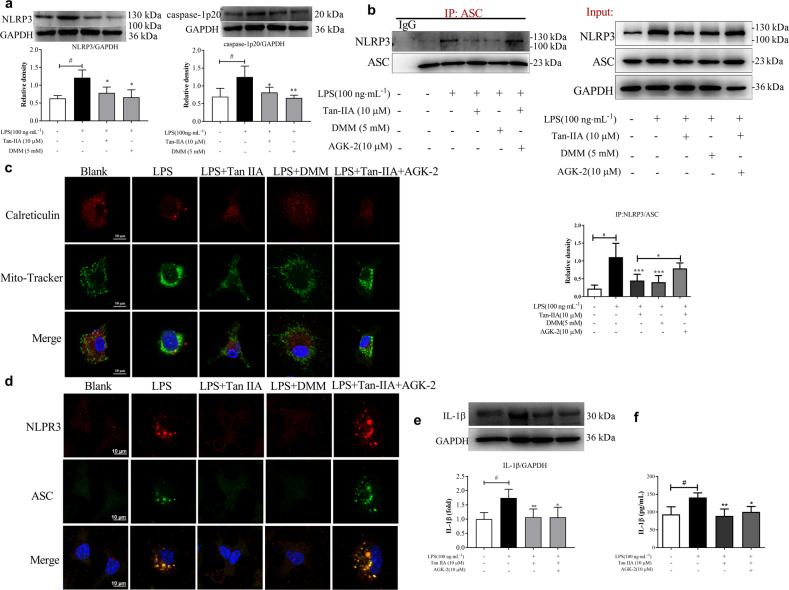
Fig. 5. Tan-IIA inactivated NLRP3 inflammasome activation through Sirt2 signaling in BMDMs.
a The protein expression of NLRP3 and caspase-1 in BMDMs stimulated with LPS (100 ng· mL−1) for 24 h; b immunoprecipitation analysis of the binding of NLRP3 to ASC in BMDMs stimulated with LPS (100 ng ·mL−1) for 24 h; c laser confocal images of the colocalization of mitochondria (stained by MitoTracker) and endoplasmic reticulum (stained by calreticulin) in BMDMs stimulated with LPS (100 ng ·mL−1) or LPS (100 ng ·mL−1) plus AGK-2 (10 μM) for 30 min (scale bar, 10 μm); d laser confocal images of NLRP3 and ASC transposition to the perinuclear space in BMDMs stimulated with LPS (100 ng ·mL−1) or LPS (100 ng ·mL−1) plus AGK-2 (10 μM) for 30 min (scale bar, 10 μm); e protein expression of IL-1β in BMDMs stimulated with LPS (100 ng· mL−1) or LPS (100 ng· mL−1) plus AGK-2 (10 μM) for 30 min; f IL-1β secretion from BMDMs stimulated with LPS (100 ng· mL−1) in the presence or absence of AGK-2 (10 μM) for 24 h (Tan-IIA tanshinone IIA, DMM dimethyl malonate). Data are expressed as the mean ± SD, n = 5. *P < 0.05, **P < 0.01, ***P < 0.001 vs. LPS-stimulated BMDMs; #P < 0.05 vs. blank.

