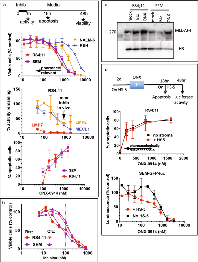
Figure 2.
MLL–AF4 cell lines are sensitive to pharmacologically relevant concentrations of ONX-0914. (a) Cells were treated with ONX-0914 for 1 h and cultured for 2 days before assessing cell viability with Alamar Blue (top panel, n = 7, SEM and RS4;11; n = 2, other), or cultured for 17 h before measuring apoptosis (bottom panel, n = 2, SEM; n = 3, RS4;11). Alternatively, cells were harvested for activity measurements immediately after 1 h treatment (middle panel, n = 2), LMP7 and LMP2 activity was measured with fluorogenic substrates, MECL1 inhibition was determined using BODIPY(TMR)-NC-005 probe. Arrows and dashed lines indicate pharmacologically relevant concentrations. (b) Cells were treated with Btz or Cfz for 1 h and cultured for 2 days before assessing cell viability with Alamar Blue (n = 2). (c) Cells were treated with 10 nM Btz or 800 nM ONX-0914 for 4 h. MLL–AF4 expression was analyzed by western using histone H3 as a loading control. Uncropped images of the blot are presented on Fig. S3. (d) RS4;11 or SEM-GFP-luc cells were cultured with HS-5 stromal cells for 48 h, treated with ONX-0914 for 1 h in the absence of HS-5, and then cultured with HS-5 cells for the times indicated. Apoptosis of RS4;11 cells was measured by flow-cytometry (top panel, n = 3). Size-based gating was used to distinguish between RS4;11 and HS-5 cells (see Fig. S4). Luciferase assay was performed to access viability of luciferase-expressing SEM-GFP-luc cells (n = 2). Bottom panel in (a) and top panel in (d) contain the same data for RS4;11 cells treated in the absence of stroma.

