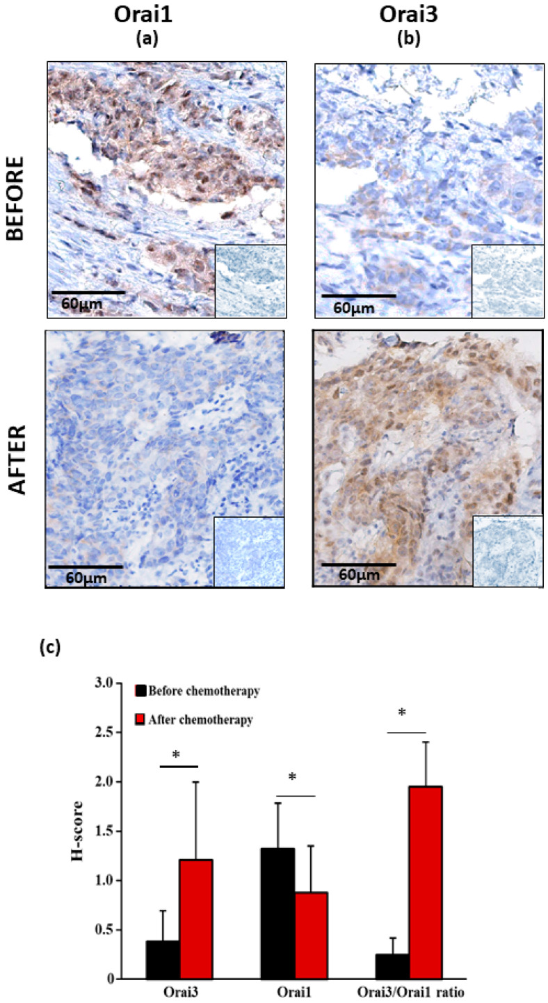
Figure 1.
Immunohistochemical staining of Orai1 and Orai3 of bronchial biopsies before and after chemotherapy. (a) Representative examples of Orai1 and Orai3 (b) expressions of original magnification: × 200. Inserts show negative controls obtained by omitting the primary antibodies. All pictures show a low magnification image of negative controls in the lower right corner. Analysis of H-score of Orai1 and Orai3 of bronchial biopsies before and after chemotherapy (c). Values are presented as mean of results obtained from 15 patients ± SEM, * p < 0.05, Mann–Whitney U test.

