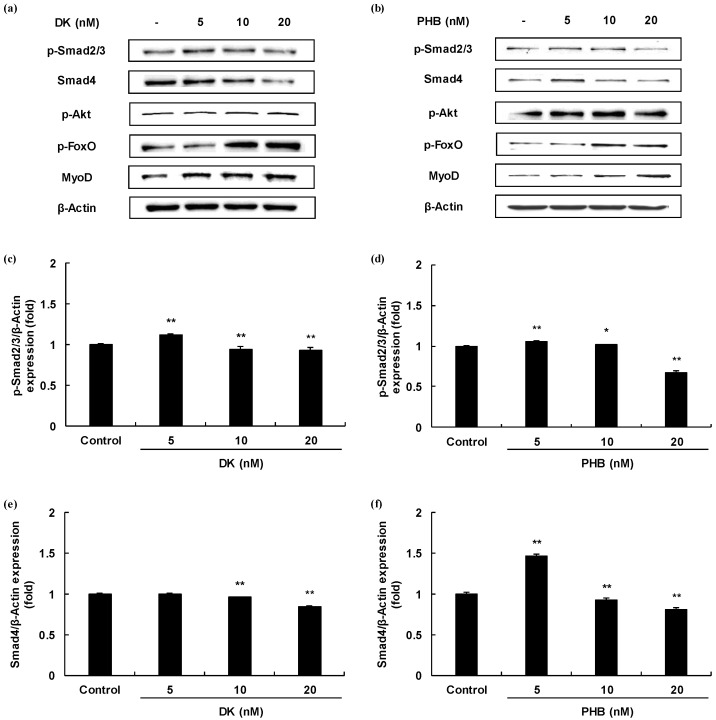
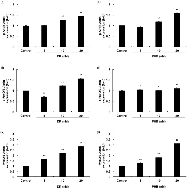
Figure 3.
Skeletal muscle cells growth activity of DK and PHB on C2C12 myotubes. (a,b), Bands of protein expressions; (c–f), TGF-β such as myostatin mediated Smad proteins expressions (p-Smad2/3, and Smad4); (g–j), IGF-1 mediated proteins expressions (p-Akt, and p-FoxO); (k,l), MyoD protein, which is known to play a critical function in the regulation of muscle cell development expressions. Experiments were performed in triplicate and the data were expressed as mean ± S.E.M.; * p < 0.05, and ** p < 0.01 as compared to the control group.


