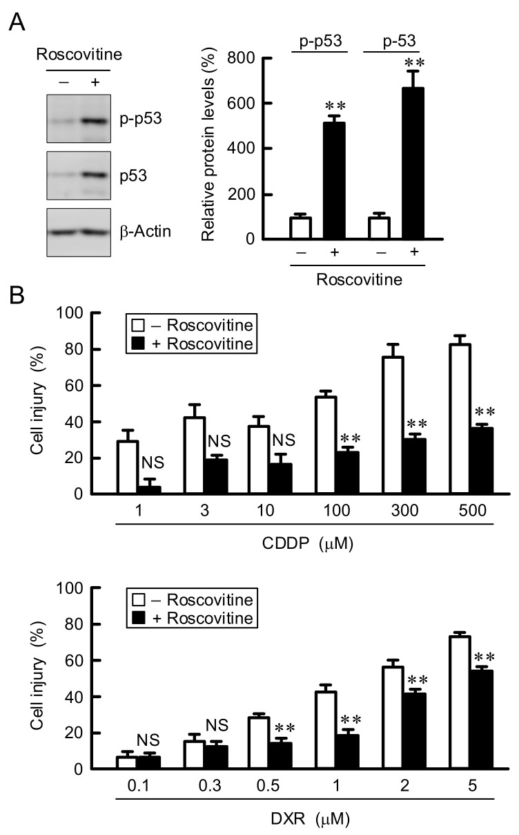
Figure 4.
Reduction of anticancer drug-induced cell injury by roscovitine. (A) A549 cells were incubated in the absence and presence of 10 μM of roscovitine for 3 h. Western blotting was performed using anti-p-p53, anti-p53, and anti-β-actin antibodies. The expression levels of p-p53 and p53 were corrected by β-actin. The protein levels are represented in percentage to the cells without roscovitine. (B) After treatment with 10 μM roscovitine for 24 h, the cells were incubated in the absence and presence of CDDP or DXR at the concentration indicated for 24 h. Cell injury was measured using the Premix WST-1 Cell Proliferation Assay System. n = 3–8. ** p < 0.01 compared with -roscovitine. NS, p > 0.05.

