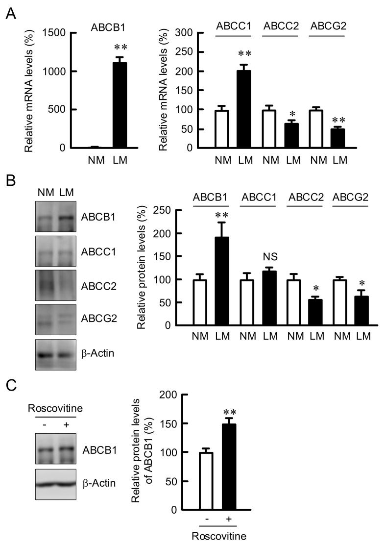
Figure 5.
Increase in the expression of ABCB1 by LM and roscovitine. (A) A549 cells were continuously cultured in the media containing NM or LM. Real-time PCR was performed using primer pairs for ABCB1, ABCC1, ABCC2, ABCG2, and β-actin. The mRNA levels are represented in percentage to NM. (B) Western blotting was performed using anti-ABCB1, anti-ABCC1, anti-ABCC2, anti-ABCG2, and anti-β-actin antibodies. The protein levels are represented in percentage to NM. (C) The cells cultured in the NM medium were incubated in the absence and presence of 10 μM roscovitine for 24 h. After Western blotting with anti-ABCB1 and anti-β-actin antibodies, the protein levels are represented in percentage to the cells without roscovitine. n = 3–4. ** p < 0.01 and * p < 0.05 compared with NM or -roscovitine. NS, p > 0.05.

