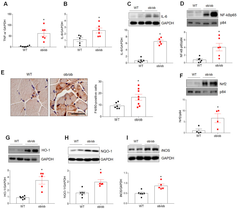
Figure 2.
Obese sarcopenia is associated with inflammation and oxidative stress in ob/ob mice. (A,B) Quantitative RT-PCR analysis of TNF-α (A) and IL-6 (B) gene expression in the skeletal muscle of wild-type (WT) and ob/ob mice. (C,D) Western blotting and quantitative analysis of IL-6 (C) and NF-κBp65 (D) expressions. (E) Representative images of immunostaining of F4/80 in cross-sections of skeletal muscle. The chart shows the number of F4/80-positive cells in F4/80-immunostained skeletal muscle sections. Scale bar = 50 μm. (F–I) Western blotting and quantitative analysis of Nrf2 (F), HO-1 (G), NQO-1 (H), and iNOS (I) expressions. GAPDH or p84 was used as an internal control to normalize total or nuclear protein levels, respectively. Data are shown as the mean ± SEM. * p < 0.05 vs. WT mice.

