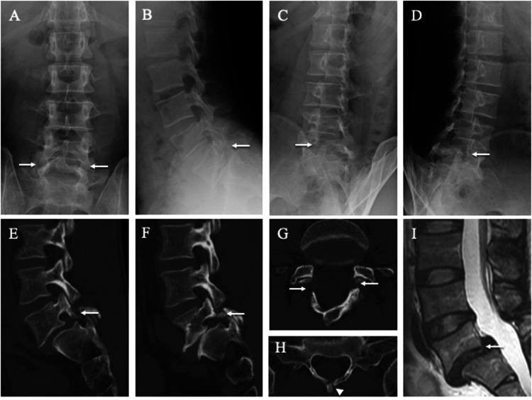
Figure 3.
Lumbar spine radiographs, computed tomography (CT), and magnetic resonance images (MRI) of the 39-year-old father. Spondylolisthesis (white arrows) is evident at the fifth lumbar vertebra (L5) on posteroanterior (a), lateral (b), 45° right anterior oblique (c), and 45° left anterior oblique (d) radiographs and right parasagittal (e), left parasagittal (f), and axial CT images parallel to the L5 (g) and first sacral (S1) (h) vertebral arches. The white arrowhead indicates spina bifida of S1 (h). L5 vertebra slippage is apparent on MRI (i).

