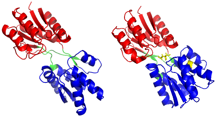
Figure 1.

(a) The X-ray crystal structure of RBP in the open-apo state (PDB code: 1URP) and (b) the closed-holostate (PDB code: 2DRI). Ribose is marked in stick representation and coloured in yellow. The C-domain of RBP is marked in red, the N-domain of RBP is marked in blue, and the hinge segments is marked in green.
