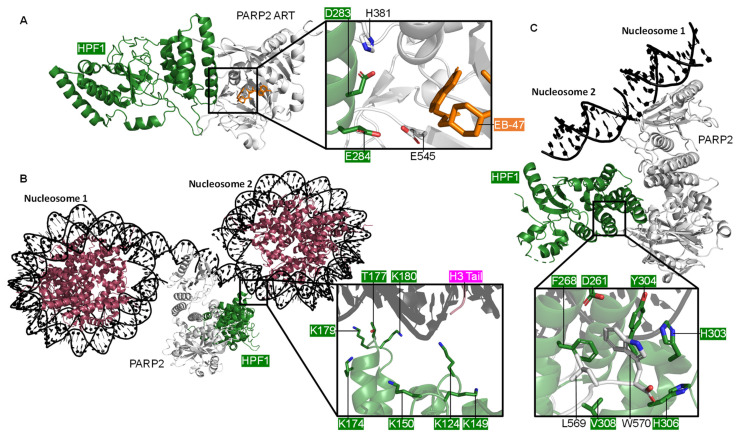
Figure 5.
The PARP2 and HPF1 Complex. (A) The crystal structure of HPF1 and PARP2 CATΔHD (ART subdomain), showing the composite catalytic site with NAD+ analogue EB-47 bound (PDB 6TX3 [150]); (B) The cryo-EM structure of PARP2 and HPF1 bridging one side of a DSB between two nucleosomes, showing the key positively charged residues of HPF1 that interact with the nucleosome (PDB 6X0N [53]); (C) A closer look at the cryo-EM structure of the PARP2, HPF1, and nucleosome complex (PDB 6X0M [53]), showing the interactions between the C-terminus of PARP2 and the C-terminal domain of HPF1.

