Figure 3.
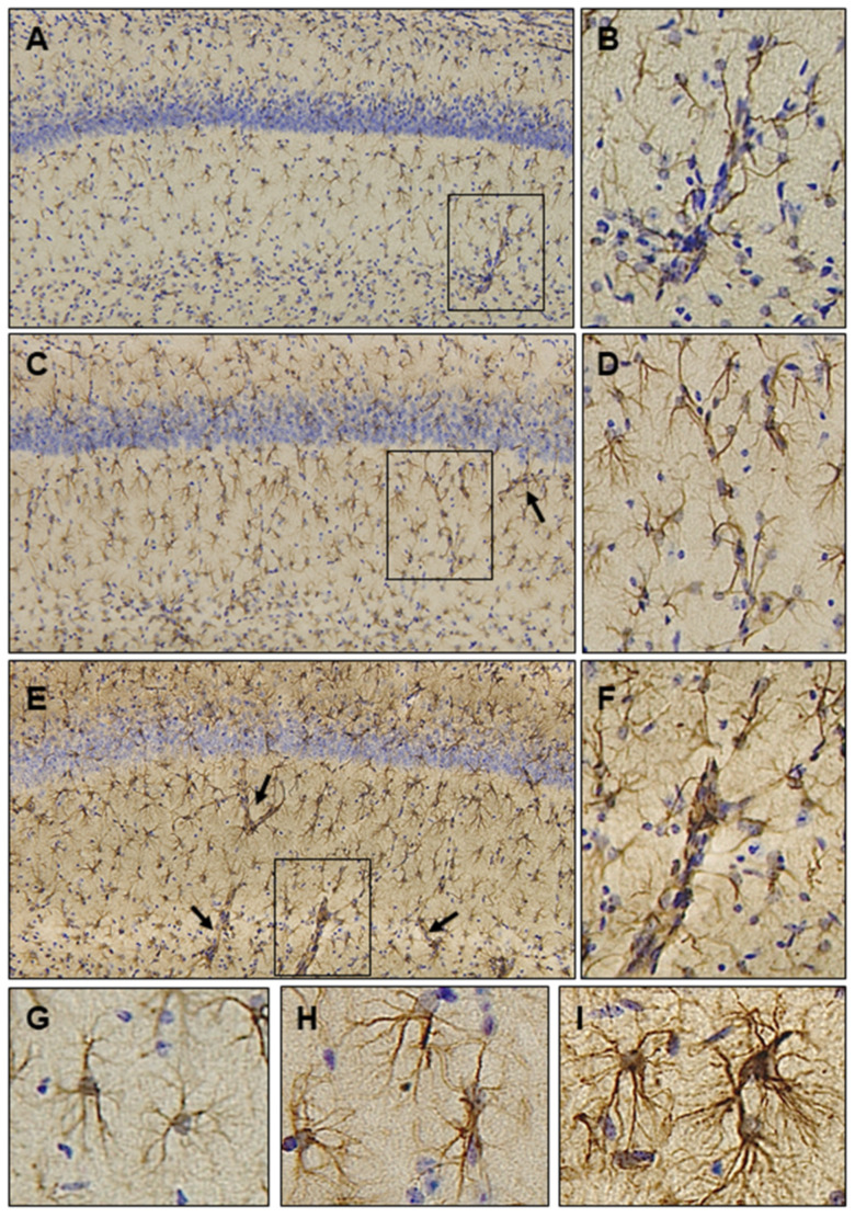
Astrocytic responses to Ubisol-Q10 supplementation in the MPTP model of Parkinson’s disease. Formalin fixed free-floating sections were subjected to immunohistochemistry using an antibody raised against glial fibrillary acidic protein (GFAP), a widely used marker of astrocytes. Astrocytes were identified by a brown precipitate at the site of antigen-antibody reaction. Nuclei were counterstained blue with hematoxylin. Shown are photomicrographs at the level of hippocampus from control (A), MPTP-injected (C) and MPTP-injected mice receiving Ubisol-Q10 (E); Magnification = 20×. Arrows in panels A, C and E depict cerebral microvasculature with increased GFAP staining forming the perivascular astrocyte endfeet. Boxed areas are represented at a higher magnification—control (B), MPTP-injected (D) and MPTP-injected mice receiving Ubisol-Q10 (F). High magnification image of activated astrocyte morphology with increased GFAP staining is shown in MPTP-injected (H) and MPTP-injected mice receiving Ubisol-Q10 (I) as compared to controls (G).

