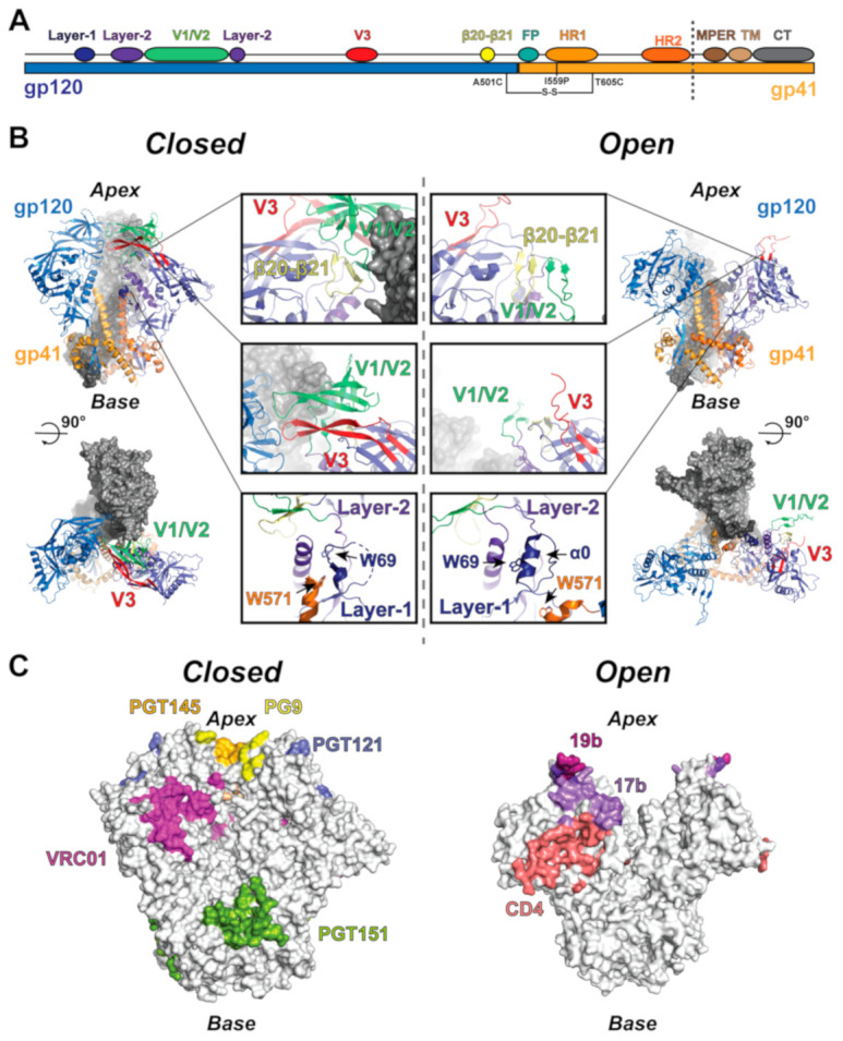
Figure 1.

Structure and allosteric elements of the HIV-1 Env trimer. (A) Linear depiction of the HIV-1 Env structural elements highlighting the position of the SOSIP mutations and the soluble ectodomain region (to dashed line). (B) (upper left) Side view of the closed state trimer. The protomer to the left is colored according to the gp120 and gp41 domains, while the protomer to the right is colored according to allosteric elements, including the β20–β21 loop (yellow), V1/V2 (lime), V3 (red), layer-1 (dark blue), and layer-2 (purple). (lower left) Top view of the closed state trimer depicting the apex gp120 contacts. (middle left) Closed state allosteric elements. (middle right) Open state allosteric elements. (top right) Side view of the open state trimer colored as the closed state trimer. (lower right) Top view of the open state trimer depicting the broken apex contacts. (C) Closed and open state surfaces highlighting the epitopes of HIV-1 Env-targeting antibodies.
