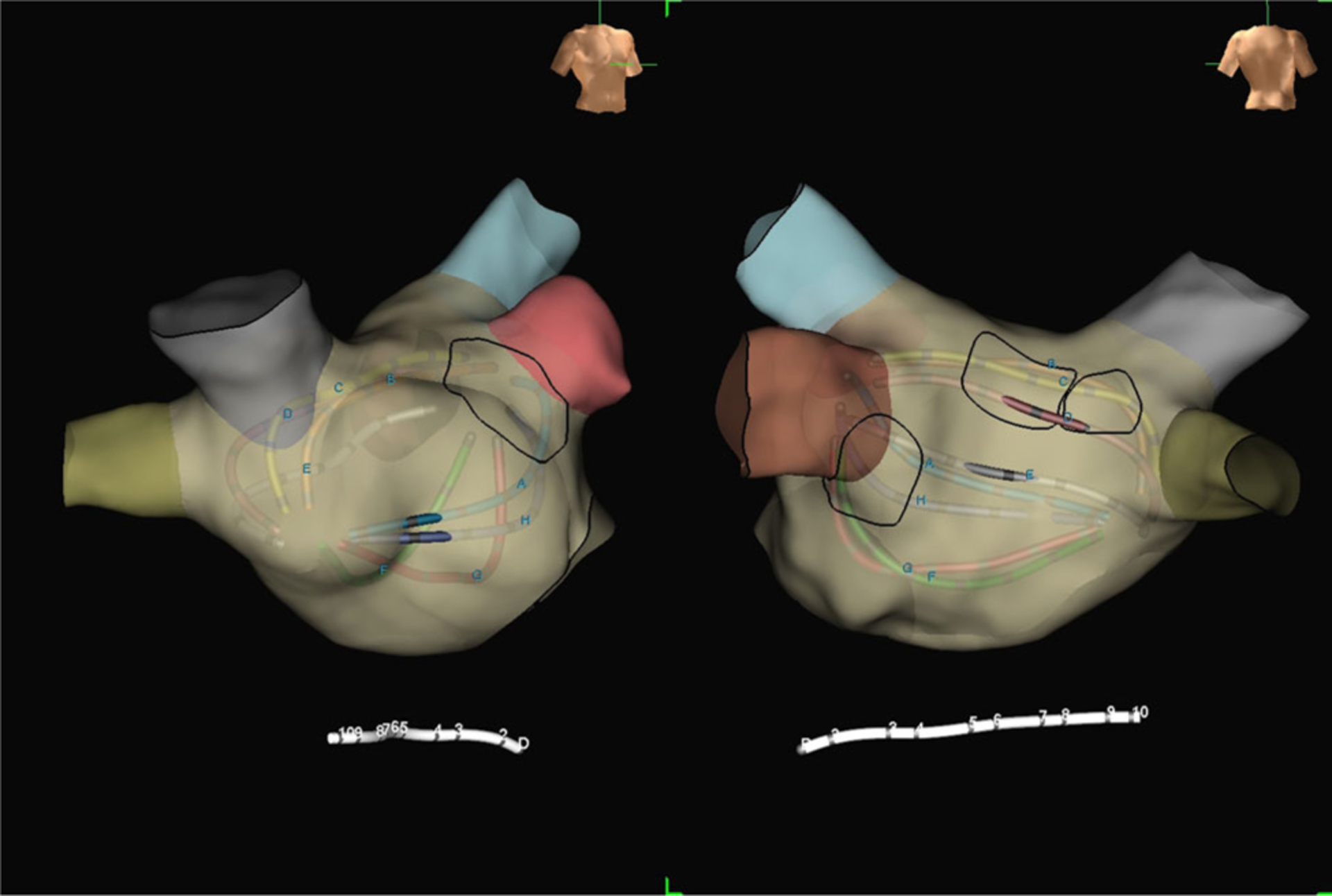
FIGURE 3.

Right anterior oblique (left) and posteroanterior (right) electroanatomic maps of the left atrium from the same 72-year-old patient undergoing FIRM plus PVRI for recurrent AF. The black circles outline three rotors (two on the midposterior left atrial wall, one adjacent to the left atrial appendage) based on mapping with FIRM software. These three rotors were subsequently targeted for ablation. AF, atrial fibrillation; FIRM, focal impulse and rotor modulation; PVRI, pulmonary vein reisolation
