
Fig. 4: Comparison of rhesus monkey and human PLD2 amino acid sequences.
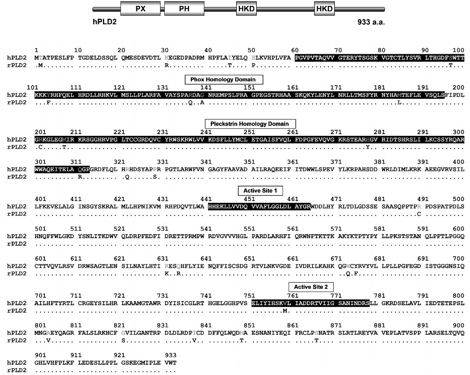
On the top is the schematic drawing of human PLD2 with its domains. PLD2 amino acid sequence from rhesus macaque was aligned with the human PLD2 protein. The human PLD2 amino acid residues are all shown. Only those residues that are not conserved in the rhesus enzyme are shown, thus, conserved residues are depicted as dots. Important protein domains are highlighted in black.