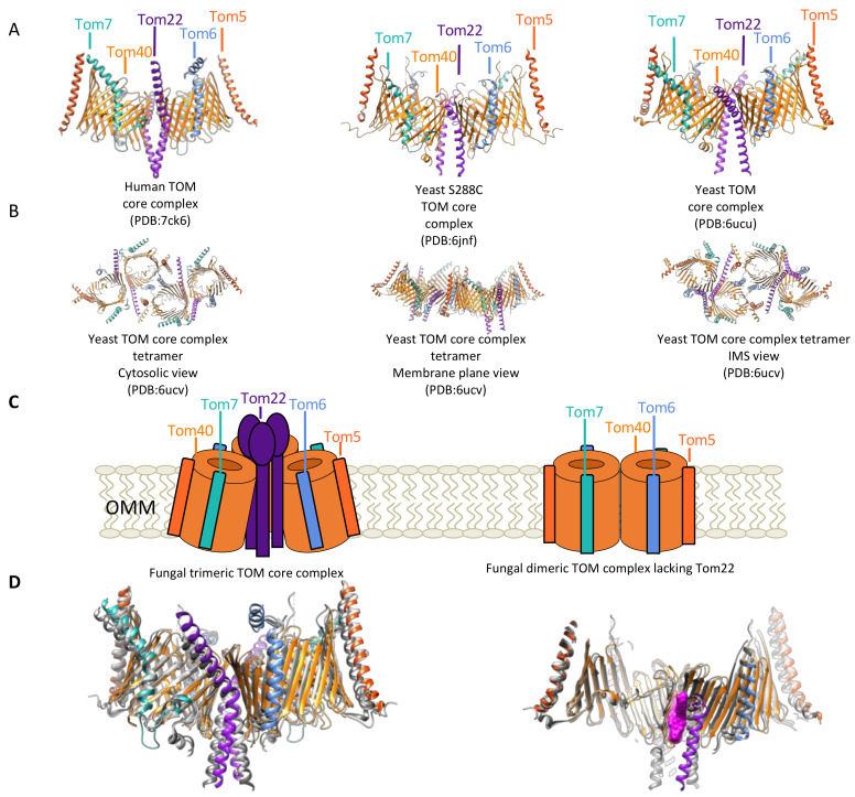
Figure 3.
Arrangements of the TOM complex. (A) Structures of the human TOM core complex (left, PDB:7ck6) illustrating high degrees of structural homology with some variations within the subunits and the dimeric form of two separately reported S. cerevisiae TOM core complex structures (middle, PDB:6jnf; right, PDB:6ucu). All three structures show two Tom40 β-barrels (orange), with two Tom22 (purple) helices between the β-barrel, opposite the Tom22 dimers on both β-barrels is a curved Tom5 (vermillion), and on opposite sides of the β-barrel between Tom5 and Tom22 are Tom6 (light blue) and Tom7 (seafoam green). (B) Structures of tetrameric yeast TOM core complex. (left) Cytosolic view of yeast TOM core complex tetramer, illustrating Tom6 role in mediating the formation of the tetramer in yeast. (middle) Membrane plane view of yeast TOM core complex illustrating the tilt of the Tom40 β-barrels and how this results in the curvature of the tetrameric TOM complex. (right) IMS view of yeast TOM core complex showing proximity of IMS domains of β-barrel associated subunits to Tom40 domains that extend into the IMS. Notably, the N-terminal region of Tom40 and Tom5, and the C-terminal region of Tom40 and Tom7 are all in close proximity. (C) (left) Cartoon representation of fungal TOM core complex trimer, (right) fungal Tom40 complex dimer lacking Tom22 based on crosslinking and biochemical data. (D) (left) Superimposition of fungal TOM complexes (gray) and human TOM complex (colored), (right) Superimposition of fungal TOM complexes (gray) and human TOM complex with lipids/detergent in magenta (space-filling model). Superimposition highlights closeness of IMS region of hTom22, extended helical region of hTom6 that runs parallel to the OMM near the cytosol, and extended loop of Tom7 near the IMS.

