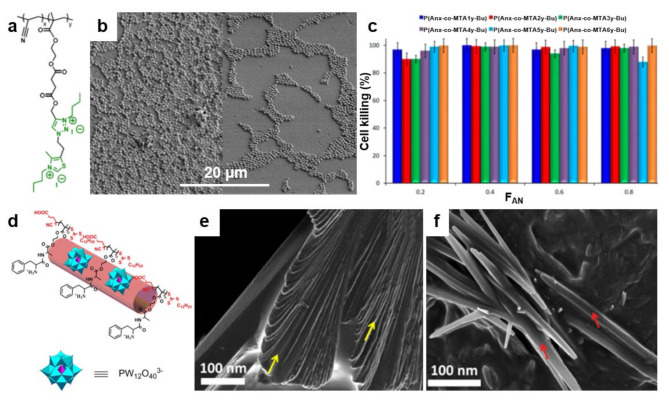
Figure 9.
(a) Chemical structure of quaternized P(AN-co-MTA) acrylonitrile-based copolymers. (b) Field emission SEM micrographs depict S. aureus on PAN films in the absence (left) and in the presence (right) of a P(AN-co-MTA) antimicrobial copolymer after 2 h of contact. (c) The percentage of cell-killing for C. parapsilosis microorganism when in contact with antimicrobial films. (d) Schematic representation of the cationic/anionic P(Boc-FA-HEMA)/POM amphiphilic supra-assemblies. (e,f) Field emission SEM micrographs depict the morphology of P(Boc-FA-HEMA) before (e) and at 90 min after the addition of POM (f). The morphology of P(Boc-FA-HEMA) is comprised of individual sheets indicated by yellow arrows in (e). These sheets dissolve after the addition of POM and lead to forming nanorods, indicated by red arrows in (f). Adapted with permission from ref. [113] (a–c) and ref. [187] (d–f).

