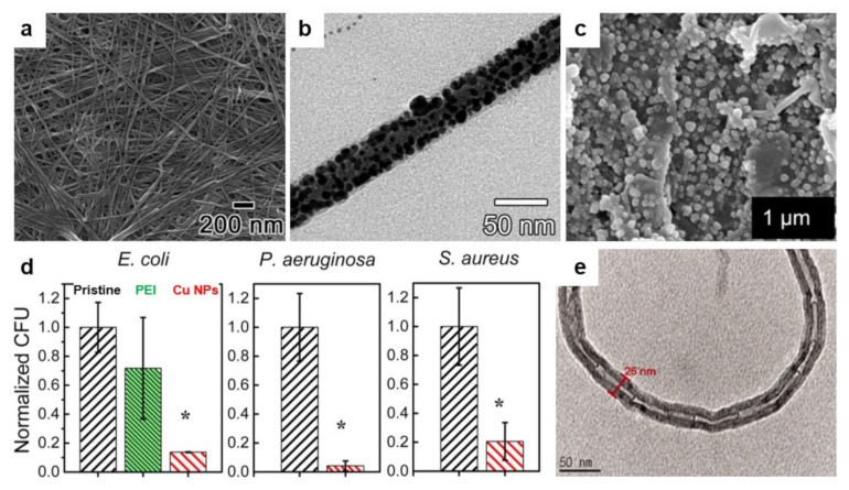
Figure 10.
(a,b) Field emission SEM (a) and TEM (b) micrographs depict biocidal Ag NPs embedded in antimicrobial PTBAM nanofibers. (c) SEM micrograph depicts a membrane with bound Cu NPs after its sonication for 5 min in deionized water. (d) Number of attached live bacteria on pristine, PEI alone, and Cu NPs-based membrane for Gram-negative E. coli and P. aeruginosa and Gram-positive S. aureus bacteria. Asterisks (*) are emphasizing statistically significant differences observed between the functionalized and pristine membranes. (e) TEM micrograph depicts the morphology of PDMEMA-MWNT nanocomposite containing almost 25 wt % of PDMEMA. Adapted with permission from ref. [190] (a,b), ref. [188] (c,d) and ref. [195] (e).

