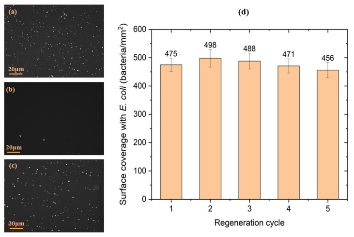
Figure 6.
Examples of microscopic images of the antibody-functionalized GaAs biochips following the initial exposure to E. coli suspension (a), after the 1st exposure to the regeneration kit (b) and after the 5th exposure to the regenerated kit followed by the exposure to E. coli (c). Evolution of the surface coverage with E. coli immunocaptured by the GaAs biochips (d) after each regeneration cycle (n = 3 samples).

