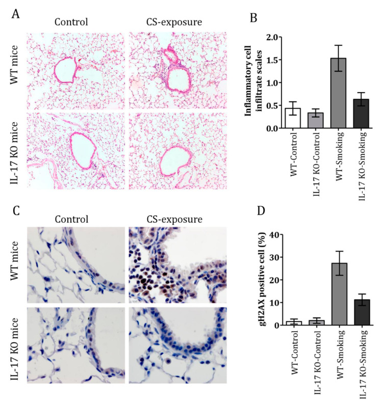
Figure 4.
Cigarette smoke induced airway inflammation and a DNA damage response in IL-17 KO mice. (A) H&E staining of lung tissue in IL-17 KO and C57BL/6 mice with or without cigarette smoke exposure (magnification 10 × 40). (B) Inflammation scores in the lungs of control and cigarette smoke-exposed mice. (C) Representative examples of gH2AX-positive cells in the lungs of IL-17 KO and control mice with or without cigarette smoke exposure (magnification 10 × 100). (D) Percentages of gH2AX-positive cells in the lungs of IL-17KO and control mice with or without cigarette smoke exposure. (n = 6 mice per group in each experiment. Data are presented as mean ± SEM).

