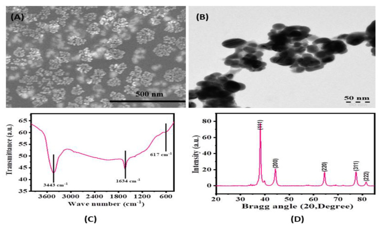
Figure 2.
(A): Scanning electron microscopy (SEM) image of biogenic silver nanoparticles (size ~15 nm), (B) Transmission electron microscopy (TEM) image of prepared silver nanoparticles from the extract (average size ~15 nm). (C) Fourier-transform infrared (FTIR) spectra of silver nanoparticles showing the functional groups in used chemicals. (D) X-ray diffraction spectrum of biosynthesized silver nanoparticles.

